
Dental Imaging
Dental imaging presents a sequence of procedures used with the aim of achieving high-definition pictures of the teeth, gum and jawbone so that the dentist can analyze what is not easily seen through naked eyes. The diagnosis of oral health issues, planning treatment, and following up should be carried out correctly.
Whether you are detecting small cavities or laying out complicated surgical pathways, dental imaging serves an unsung hero of modern dentistry. Ordinary X-rays have now transformed into sophisticated digital scans, 3D scans, and even AI-based diagnostics, which ensure quicker and safer dental care and are much more accurate than they have ever been.
Jamaica 26 Dentistry implements the latest technology in dental imaging techniques to provide the most precise and personalized care for every smile.
Importance of Dental Imaging
However, dental imaging is deeper than a one-day X-ray. It is the backbone of efficient, informed, and personal dental care. The following are the points that make these techniques important:
Early Detection of Oral Issues:
Imaging can identify issues before they develop into bigger problems, everything else goes from small holes in between teeth to initial evidence of bone loss, infection, or abscess. As the problems are diagnosed earlier, they become more manageable and less painful to treat.
Accurate Diagnosis
A visual examination is limited. Imaging provides a complete inside view that ensures that your dentist can correctly diagnose complex conditions such as impacted teeth, jaw misalignment and concealed decay.
Essential for Treatment Planning
Whether it is dental implants, root canals, or braces, imaging is essential in mapping the treatment plan. It adds precision to everything, reducing risk and increasing the success rate.
Preventive Care & Long-Term Monitoring
Periodic X-rays are useful in monitoring dental health. It acts as a medical history of your teeth, which will help you check any changes, pick up the same problems and offer special care to prevent dental problems.
At Jamaica 26 Dentistry, precision diagnosis, customized treatment plans and proactive ways of caring are combined with top-quality and trained dentistry personnel, state-of-the-art imaging leaving your smile healthy, strong and stress free.
Common Types of Dental Imaging
Learning the variations of dental imaging can help patients understand the role each method plays in diagnosis and treatment planning. Here is an analysis of the most popular methods:
Intraoral X-rays
They are the most prevalent form of dental imaging and offer exceptionally detailed glimpses of particular sections within the mouth.
- Bitewing X-rays are utilized to visualize upper and lower teeth in a single region revealing decay between teeth and bone levels.
- Periapical radiography is an x-ray that takes all the parts of the tooth, including the crown and roots, to detect abscesses, cysts, or root problems.
- Occlusal X-rays provide a wider picture of the floor or roof of the mouth to identify impacted teeth and fractures of the jaw.
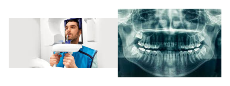
Extraoral X-rays
They were used to absorb more significant structures without the teeth, which included the jaw and skull.
- Panoramic X-rays are presented as one image reflecting the entirety of the upper and lower jaw, making them ideal to use when examining wisdom teeth, diseases of the jaw, and planning implants.
- Cephalometric projections are mainly used in orthodontics to analyze tooth displacement and jaw development.
Cone Beam Computed Tomography (CBCT)
It is a specialized 3D image that is used to provide detailed cross-sections of the oral and maxillofacial structures.
- This imaging technique is essential to measure bone density, nerve pathway, and location of sinus frequently utilized in preparing implants, treating root canal and during surgeries.
Digital Radiography
The modern form of traditional X-rays utilizes digital sensors rather than film.
- It provides quicker processing of images, less radiation exposure, and facilitates storage and sharing of patient records.
Intraoral Cameras
High resolution cameras take live images of the mouth.
- Appropriate to identify cracks, plaque deposits, and other surface-related problems. Also useful in informing patients of their oral health status.
MRI and CT Scans (in complex cases)
These are not used regularly in general dentistry, Magnetic Resonance Imaging (MRI), and conventional Computed Tomography (CT) scans could be necessary in diagnosing diseases of the temporomandibular joint (TMJ) disorders, or in assessing conditions of the soft tissues or extensive facial trauma.
Advantages of Modern Dental Imaging
With the evolution in technological applications in the dental sector, the new technologies of imaging have introduced significant changes in the diagnosis and management of oral health issues. These developments are linked to a wide range of benefits that augment patient experience and clinical results:
- Reduced Radiation Exposure
Digital imaging technology uses significantly less radiation than outdated film recordings of X-rays. This produces no risk to patients but does not degrade image quality, which is critical in the case of scanning children, pregnant individuals, or patients that require repetitive scans. - Faster Diagnosis
Digital imaging response in real-time and permits the dentist to analyze and examine in a real-time environment. This reduces waiting time, physical film development and allows the treatment decision to be made quickly. - Better Treatment Planning
Images must be high-resolution to allow more detailed treatment planning, especially in more complicated cases, e.g., inserting implants, root canal treatment, and orthodontics. Dentists are able to examine the structure of bones, teeth alignment, and nerve location with stringent accuracy. - Improved Patient Communication
The use of visuals is effective. Using intraoral cameras and digital imaging, the patient sees what is going on in his/her mouth. This creates a level of trust, understanding and patients become more active decision-makers in their treatment. - Digital Storage & Easy Sharing
Photographs are kept in a digital format, which is more efficient in terms of record-keeping. They are easily accessed for follow-ups, referred to specialists, and shared with companies handling the insurance, all without the inconvenience of a physical file of delays.
Safety and Radiation Concerns
- Dental imaging used today is associated with very low doses of radiation.
- Compared to the film X-rays, digital X-rays produce low radiation.
- Sensitive areas are covered with lead aprons and thyroid collars.
- Precise safety measures are taken to protect the patient.
- The principle of ALARA (As Low As Reasonably Achievable) is used at all times.
- Children, pregnant patients (when required), undergo imaging with utmost care.
When Do You Need Dental Imaging?
Dental Imaging not only in an emergency but is also critical in ensuring long-term oral health. These are the most typical cases when it is required:
Routine Check-Ups
Your mouth might feel good but frequent dental X-rays help detect any problems you might not be aware of during routine checkups. They can diagnose early tooth deterioration, removal or prevent infections before they become worse, making treatment less invasive and simple.
When Experiencing Pain or Swelling
A common sign of a possible problem is chronic tooth pain, gum pain, or jaw pain. Dental imaging will allow your dentist to diagnose which cause is present, e.g. an abscess, impacted tooth, cyst, or jaw infection and provide early treatment at that time.
Before Dental Procedures
Imaging is also necessary when receiving a crown, dental implant, orthodontic work, or oral surgery. It offers complete details concerning the structure of the bones, placement of teeth, and the location of the nerves to give safe, exact and successful results.
For Monitoring Chronic Conditions
Patients with constant tooth issues such as patients with periodontitis, jaw seeds, or even patients with a history of frequent cavities can be imaged regularly. These scans may be used to help track the disease’s progress, response to medication and make timely adjustments to prevent any injury.
The Future of Dental Imaging:
Advanced technologies are shaping the future of dental imaging, even rendering it faster, accurate, and highly customized. Artificial intelligence (AI) is turning into a gamechanger that enables dentists to detect even subtle abnormalities on the scans, make the diagnosis more accurate, and reduce human error.
The integration of 3D printing and imaging can help dentists create highly precise surgical guides, custom prosthetics, and orthodontic devices in order to ensure that each patient receives a personalized treatment that takes into account their specific anatomy.
The use of teledentistry and remote diagnostics is increasing the accessibility of expert dental care since dentists can now access imaging results, patient consultations and treatment planning without a person having to visit their premises. These innovations are all transforming dental imaging into a smarter, faster and more patient-centered use of oral healthcare.
FAQs
Is dental imaging safe during pregnancy?
Yes, in most cases dental imaging is safe during pregnancy particularly when we have high-quality digital X-rays with a modest amount of radiation. But, to be on the safe side, imaging is usually avoided in the first trimester unless there is no other option. Both mother and baby are shielded using lead aprons and thyroid collars when necessary. Inform your dentist about your pregnancy so that he/she takes extra measures.
How often should dental X-rays be taken?
The frequency of dental X-rays depends on the health status of your mouth, your age, risk factors, and previous dental issues. In adults with no recent complications, X-rays can be prescribed every 12-24 months. The dentist can assess whether they are more likely to be needed by chronic dental subjects, children, or those undergoing treatment.
What’s the difference between panoramic and bitewing X-rays?
When imaging the entire upper and lower jaws, teeth, sinuses, and jaw joints in a single image, panoramic X-rays are especially useful in evaluating impacted teeth, assessing jaw growth and development, or planning dental implants. Bitewing X-rays, focus on the upper and lower back teeth in the same area and are mainly used to detect dental caries between teeth and monitor bone levels.
Do I need to prepare for a dental scan?
Dental imaging procedures can be done without any special preparation in most cases. Your jewelry and glasses may also be requested to be removed prior to the scan. It will take a couple of minutes and only a little pain if you are going in for a CBCT scan or panoramic X-ray, these procedures are done based on the instructions created by the dentist or technician in charge.
Can children undergo dental imaging?
Absolutely. Dental imaging is both safe and typically needed by children to observe the maturation of teeth, diagnose decay early, and prepare orthodontic treatment. Lower doses of radiation are used in pediatric imaging, and all typical precautions are taken to protect children.